What Is Grade 1 Maturity In Ultrasound Scanning
Her Ultrasound pregnancy level II reports show the following. A single live intrauterine fetus is seen in breech presentation at the time of scanning.

Ageing Or Calcification Of The Placenta Babycenter India
Abruptio Placentae C0000832 Definition NCI Placental separation from the uterus with bleeding concealed or vaginal before fetal birth with or without.
What is grade 1 maturity in ultrasound scanning. Grade 2 is mature. Subsequently one may also ask what do you mean by Grade 3 placenta. Placenta is anterior of grade 1 maturity.
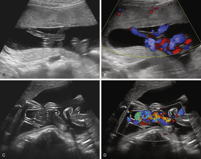
It is normally closed in the antenatal period and opens only at the time of labor. Gianoukakis MD FACE. 2- Color Doppler reveals non-fillilng of external lilac vein on the left side.
The placental grading will be given according to its. It is called migration of placenta. Maturation is the process of achieving full development or growth.
In some countries the use of placental grading has fallen out of obstetric practice due to a weak correlation with adverse perinatal outcome 5. 1- grey scale imaging shows enlarge diameter of Common Femoral Vein CFV Superficial Fomoral Vein SFV. XXXXXXX os is.
Grade 1 maturity What is ultrasound scanning. 123mmcorresponding to 18 weeks 4 day and FL. It is normal grading of placenta.
This primarily affects the extent of calcifications. The mean gestational age at which the placenta matures to grade 1 is 31 weeks grade 2 is 36 weeks and grade 3 is 38 weeks as seen by others as well. Placental substance - Few scattered echogenic areas appear within the placenta resulting in a loss of homogeneity.
Hi As per your sonogram report everything seems to be normal and correlating to your gestational age except that the placenta is low-lying. The Graf method for ultrasound classification system for developmental dysplasia of the hip DDH in infants combines both alpha and beta angles. There is no retroplacental collection.
Grade 3 is hyper mature. The problem depends on gestational age. 2 Single loop of cord around fetal neck is seen in.
I would suggest stk to ask her doctor cos usually by 19 weeks my placenta was grade 0 in first pregnancy and grade 1 in second. HC145mmcorresponding to 17weeks 5 day. Mismatched grades for gestational age are considered abnormal.
Doctor my mother aged 63yrs have DVT. Grading is between 0 and 3 with 3 being the mature placenta at 40 week Ontology. Ultrasound technology uses sound waves to develop images of body composition.
It is said that maturation of the placenta does not occur at the same rate and same degree in all pregnancies. It is a homogenous echopattern grade 1. 24mmcorresponding to 17 weeks 3 day.
There are a number of additional subdivisions which are often not used clinically. Rakhi Tayal OBGYN 20 weeks pregnant. Grade - 1 It is the earliest ultrasound changes of placental maturation.
I dont think its a matter of concern until placenta shows grade 4 maturity but ask ur doc for clarification. Ultrasound scanning for carcass traits is a useful tool for obtaining valuable carcass information from a live animal. Fetal heart rate is regular and is 157 beats minute.
The inferior limit extends down to the XXXXXXX os but does not span across it. 1 Your baby is mature enough with normal weight and corresponds to current gestational age. Usually in early gestational age the placenta could be some what low lying and with progression of gestational age it will move upwards.
The scanning which is done by using ultra sounds is called as ultra sound scanninglikescanning the. Click to see full answer. Placental grading Grannum classification refers to an ultrasound grading system of the placenta based on its maturity.
Thyroid Ultrasound Basic ATA Fellow Track 2013 LABioMed Andrew G. Placenta is also mature and placed in right position. Grade B BEL 3.
5cm scanning depth The neck is scanned in both transverse and longitudinal planes. 40mmcorresponding to 18 weeks 1 day. Nalinithats grade for placenta previanot placenta maturity.
An M-mode recording is conventionally displayed with the abscissa representing time and the ordinate distance from the. First of all I want to explain your ultrasound scan report one by one. Fetal movements are good.
M-mode ultrasound Often utilised for its excellent axial and temporal resolution of structures M-mode or motion mode is a form of ultrasonography in which a single scan line is emitted received and displayed graphically. Placental maturity may be accelerated or delayed in certain conditions. Chorionic plate - Well-defined unbroken line but may present fine undulation.
The placenta is anterior. The mean gestational age at which the placenta matures to grade 1 is 31 weeks grade 2 is 36 weeks and grade 3 is 38 weeks as seen by others as well. Body composition traits that can be measured include 12th to 13th rib fat thickness rump fat thickness ribeye area and intramuscular fat percentage marbling.
As a general rule the alpha angle determines the type and in some instances the beta angle is used to determine subtype. Os is the opening in the cervix through which the baby descends during labor. When scanned the report says following.
Thyroid Ultrasonography Normal Anatomy. Likewise what is maturity in pregnancy. You should be assessed by your obstetrician.
Ultrasound showed anterior placenta. Lower limit of placenta is not reaching the XXXXXXX os. Placenta Fundal Posterior Grade 1 maturity means the Placenta is located towards the back of the uterus and Grade 1 shows the maturity of the Placenta.
Partial filling of colour is seen in the left CFV SFV. The ultrasound result for my expecting wife at 17 weeks and six days is as follows. Hi Thanks for the query.
Grade 1 is immature placenta only.

Marginal Placenta Previa Sonosession

Pregnancy Week 35

Third Trimester Growth Scan Babycenter India

Fetal Lung Maturity Assessment A Historic Perspective And Non Invasive Assessment Using An Automatic Quantitative Ultrasound Analysis A Potentially Useful Clinical Tool European Journal Of Obstetrics And Gynecology And Reproductive Biology

Pin By Emily Kuske On Ob Ultrasound Obstetrics Gynecology Uterus

Outcomes Of Pregnancies With A Low Lying Placenta Diagnosed On Second Trimester Sonography Heller 2014 Journal Of Ultrasound In Medicine Wiley Online Library

Ultrasound Imaging Of The Placenta Part 1 Anatomy And Variants Youtube
Scan Of The Week 20 Week Detailed Anomaly Scan Youtube
Https Www Glowm Com Pdf Ultrasound In Obstetrics And Gynecology Chapter10 Pdf
Ultrasound Evaluation Of The Placenta Membranes And Umbilical Cord Radiology Key
![]()
Ultrasound Scan At 30 Weeks Of Gestation Cystic Appearance Of Molar Download Scientific Diagram

Week 36 Ultrasound What It Would Look Like Parents
Calcification Ageing Of Placenta During Pregnancy

Exploring The Relationship Between Preterm Placental Calcification And Adverse Maternal And Fetal Outcome Chen 2011 Ultrasound In Obstetrics Amp Gynecology Wiley Online Library
Week 27 Ultrasound What It Would Look Like Parents

Pelvic Ultrasound Scan Of A 26 Year Old Pregnant Patient With Ovarian Download Scientific Diagram

Marginal Placenta Previa Sonosession

What Is A Placental Lake Babycentre Uk

Does Your Anomaly Scan Say You Have Posterior Placenta Here Is What It Means Thehealthsite Com